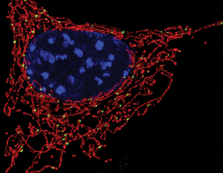
One of the earliest detectable signs of retinal dysfunction – before even anatomical changes can be seen – is mitochondrial dysfunction (1), (2). That’s understandable; photoreceptors – both rod and cones – are highly metabolically active, with (ironically enough) rods being most active during sleep (3). It’s becoming clear that because these cells have such a high metabolic rate, it makes them especially vulnerable to mitochondrial dysfunction.
But just how vulnerable are these cells? To determine that, researchers need a direct, reliable and easy-to-use assay of mitochondrial function – and that’s exactly what a team of researchers from the (US) National Institute of Health have produced; a microplate-based method that enables a high-throughput assay of the oxygen consumption rate (OCR) of mitochondrial cells in acutely isolated, ex vivo mouse retina (4). What they found was that photoreceptor mitochondria have very little in the way of reserve capacity to respond to stress – they work close to their maximum, even at a baseline state. It has previously been suggested that early alterations to the mitochondrial reserve capacity can predict later photoreceptor cell death (5), and this work by the NIH team reinforces and confirms that viewpoint, suggesting that the use of mitochondrial OCR can be used as an experimental early biomarker of retinal dysfunction. These findings help to explain why the retina is so sensitive to even relatively small changes to energy homeostasis that are caused by mutations or metabolic challenges. “We show for the first time that mitochondria within photoreceptors operate at 70 to 80 percent of their maximum capacity, with very little reserve, which suggests that the cells are in a perpetual state of high metabolic stress. It’s like when a rubber band gets stretched all the way to the point where adding just a little more force breaks it. Our data strongly support the use of mitochondrial oxygen consumption rate as an early biomarker of retinal disease, before the onset of overt degeneration,” says Anand Swaroop, lead author of the associated paper (3).
References
- R McGuigan, “Light years ahead”, The Ophthalmologist, 22 (2015). Available at: http://bit.ly/TOPLight M Barot et al., “Mitochondrial dysfunction in retinal diseases”, Curr Eye Res, 36, 1069–1077 (2011). PMID: 21978133. I Grierson, R Kirk, “Nocturnal light saves sight”, The Ophthalmologist, 25 (2015). Available at: http://bit.ly/TOPNLSS. K Koorgayala et al., “Quantification of oxygen consumption in retina ex vivo demonstrates limited reserve capacity of photoreceptor mitochondria”, Invest Ophthalmol Vis Sci, 56, 8428–8436 (2015). PMID: 26747773. NR Perron et al., “Early alterations in mitochondrial reserve capacity; a means to predict subsequent photoreceptor cell death”, J Bioenerg Biomembr, 45, 101–109 (2013). PMID: 23090843.
