Conexiant
Login
  • Corneal Physician
  • Glaucoma Physician
  • New Retinal Physician
  • Ophthalmology Management
  • Ophthalmic Professional
  • Presbyopia Physician
  • Retinal Physician
The Ophthalmologist
  • Explore

    Explore

    • Latest
    • Insights
    • Case Studies
    • Opinion & Personal Narratives
    • Research & Innovations
    • Product Profiles

    Featured Topics

    • Anterior Segment
    • Glaucoma
    • Retina

    Issues

    • Latest Issue
    • Archive
  • Subspecialties
    • Cataract
    • Cornea
    • Glaucoma
    • Neuro-ophthalmology
    • Oculoplastics
    • Optometry
    • Pediatric
    • Retina
  • Business

    Business & Profession

    • Professional Development
    • Business and Entrepreneurship
    • Practice Management
    • Health Economics & Policy
  • Training & Education

    Career Development

    • Professional Development
    • Career Pathways

    Events

    • Webinars
    • Live Events
  • Events
    • Live Events
    • Webinars
  • Community

    People & Profiles

    • Power List
    • Voices in the Community
    • Authors & Contributors
  • Multimedia
    • Video
    • Podcasts
Subscribe
Subscribe

False

Advertisement
The Ophthalmologist / Issues / 2021 / Dec / No Noise Is Good News
Retina Business and Entrepreneurship Practice Management Sponsored

No Noise Is Good News

Using deep learning to denoise B-scan OCT images

Sponsored By Nidek 12/10/2021 1 min read

Share

a. Single-frame image (left). b. After denoising (right). Images courtesy of Rodrigo Abreu González, MD, PhD, FEBO, University Hospital of La Candelaria, Spain.

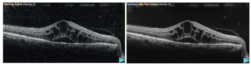
Optical coherence tomography (OCT) has become the most popular and powerful retinal imaging system in the ophthalmology market. Nevertheless, as with any other modality, the images it produces are subject to noise – either from the patient’s eye movements, media opacities (such as cataracts, blood, or dense vitreous), or technical factors (such as slow scanning speed). OCT noise can be a real problem for clinicians – and overcoming it without increasing capturing time is hugely beneficial.

In October 2021, NIDEK, a renowned manufacturer of ophthalmic equipment, launched the Retina Scan DuoTM2 – a combined OCT and fundus camera system, incorporating B-scan Denoising Software for quick acquisition of high-definition OCT images. The new method has the capability of converting a single B-scan retinal OCT image into a high-definition image, thanks to a deep-learning based denoising technique. Notably, the software is also available for other NIDEK OCT devices, such as the Mirante and the RS-3000 Advance 2. 

Different from image averaging – a standard process used to create clear B-scan images – NIDEK’s unique denoising technique does not require the capture of multi-frame images to remove speckle noise.

a. Single-frame image (left). b. After denoising (right). Images courtesy of Rodrigo Abreu González, MD, PhD, FEBO, University Hospital of La Candelaria, Spain.

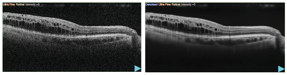
How does the denoising software work – and how was it created? First, NIDEK adopted an effective deep-learning platform (a generative adversarial network) known to improve image clarity. Next, a large number of images (each one averaged from 120 B-scan frames) was used to train the deep learning platform how to create images from a single frame that are comparable in clarity to those generated by averaging 120 images. As a result, when using the software with the NIDEK OCT, once a B-scan image is captured, a denoised version is automatically displayed, without the need for longer capturing time.

Acquiring high-quality images rapidly is extremely valuable for both physicians and patients, as it results in better patient flow and increased patient comfort. Providing exquisite image quality, this software is particularly useful when capturing multiple B-scan images for wide area screening – truly a new solution to overcome OCT speckle noise. 

Please note that registration for this software is not yet available in China. The availability of the OCT devices mentioned here differs from country to country, depending on the approval status.

Related Content

Newsletters

Receive the latest Ophthalmology news, personalities, education, and career development – weekly to your inbox.

Newsletter Signup Image

False

Advertisement

False

Advertisement

Explore More in Ophthalmology

Dive deeper into the world of Ophthalmology. Explore the latest articles, case studies, expert insights, and groundbreaking research.

False

Advertisement
The Ophthalmologist
Subscribe

About

  • About Us
  • Work at Conexiant Europe
  • Terms and Conditions
  • Privacy Policy
  • Advertise With Us
  • Contact Us

Copyright © 2025 Texere Publishing Limited (trading as Conexiant), with registered number 08113419 whose registered office is at Booths No. 1, Booths Park, Chelford Road, Knutsford, England, WA16 8GS.

Disclaimer

The Ophthalmologist website is intended solely for the eyes of healthcare professionals. Please confirm below: